by Lisa Davis
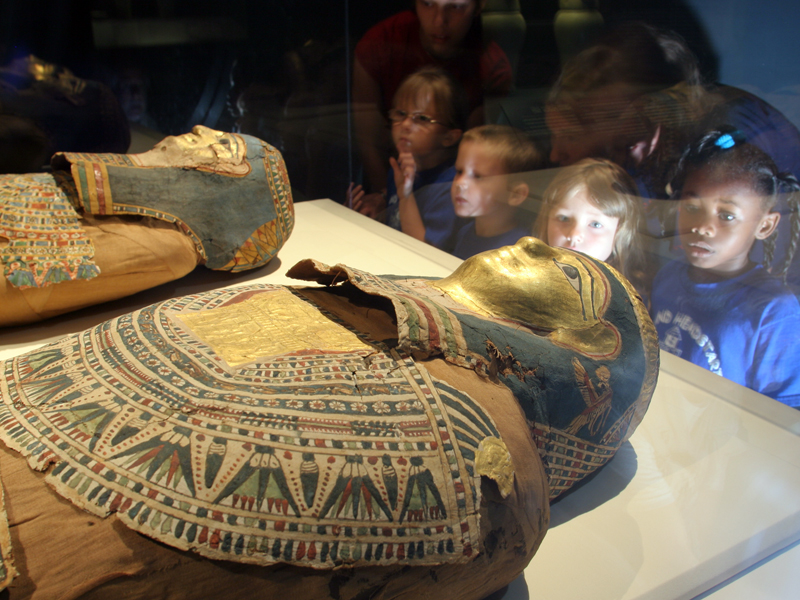
For years, we’ve known her name, spelled out in hieroglyphs on the painted paper covering her body: “Tasherytpamenekh, justified of voice forever and ever.”
It’s pronounced Ta-SHER-eet-pa-MEN-eck, and it means “daughter of Pamenekh.”
Now we know even more about the Anniston Museum of Natural History’s mummy, thanks to images released this week from a high-tech CT scan at Regional Medical Center.
The scans, according to Dr. Robert Garner of RMC, revealed a remarkably well-preserved skeleton for a 2,300-year-old woman.
She had strong bones and good teeth, signs that she ate well, and was probably in her 20s. She might have been wealthy; her joints show none of the stress that would have marked her as a laborer.
She has no lungs, or stomach, or liver. Her organs were taken out during the mummification process. (Her brain was removed by sticking a long hook up her nose.) Even if her organs remained, Garner said they would be unrecognizable now.
There was no jewelry on her body. There might never have been, or it might have been stolen by grave robbers.
The big mystery, though, remains: What killed her?
“There are no fractures, nothing to suggest a traumatic death, or to suggest a hard life,” Garner said.
“At that time, there were plagues going through. She could have been on the wrong side of a political struggle. She could have been poisoned,” he added. None of those means of death would leave evidence we could read today.
Arrival in the U.S.
The Anniston Museum’s two Egyptian mummies first appeared in America in 1926. A dealer in Cairo hoped to sell them at the Sesquicentennial Exhibition in Philadelphia, Pa., but U.S. Customs wouldn’t release the mummies.
A man named H. Severn Regar, who had a museum in Norristown, Pa., visited the exhibition and heard about the mummies, and arranged to buy them for his museum. Regar later moved to Anniston, bringing with him the mummies and the rest of his artifacts — notably a collection of mounted birds. He donated his collection to the city for a museum.
X-ray vision
This is not the first time the mummies have been examined medically. In 1978, they were X-rayed before being sent to New York for preservation work. Those X-rays revealed that the smaller of the two mummies was a young woman, with a bit of arthritis in her spine and neck. At the time, they assumed she was in her late 30s.
The larger mummy was in much worse shape, with a crushed pelvis, lower spine and two broken ankles — damage that may have been the cause of death, or the result of grave robbers.
Mummy or daddy?
The story has always been that the mummies were mother and daughter. But in X-rays taken in 1978, the larger mummy’s pelvis turned out to be so badly damaged, it’s impossible to tell if it is male or female.
King Tut’s scan
The body of King Tut received a high-tech CT scan in 2005, which revealed that he was not killed by a blow to the head, or by political assassination, as previously thought. The scans did reveal a broken leg, which quite possibly got infected and led to the boy king’s untimely death.
When they lived
The two mummies lived during the Ptolemaic Era, c. 332-30 B.C. The Greeks, led by Alexander the Great, conquered and ruled Egypt. The Greeks established the city of Alexandria, and its great museum and library. The Ptolemaic era ended with the death of Queen Cleopatra and the Roman conquest of Egypt.
Where they lived
The two mummies are from the ancient Egyptian city of Thebes, the capitol of Upper Egypt, located on the Nile River. Thebes is home to the Valley of the Kings, where tombs for the pharaohs were built for nearly 500 years. The modern-day cities of Luxor and Karnak are near there.
How mummies were made
The ancient Egyptians made mummies because they believed they would need their bodies in the afterlife. So they invented a way to preserve them.
During mummification, the internal organs were removed and preserved in separate sculpted jars. Wine was used to wash the body. Spices were used in embalming the body.
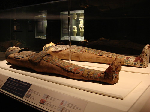
The mummy’s clothes
After embalming, a body was wrapped in strips of linen. These linen strips are visible in the CT scans. There’s also a box of mummy wrappings back in the museum’s storage area. They fell off during the 1978 restoration work. Few people in the museum will touch the wrappings with their bare hands, because they’re superstitious about the mummy’s curse.
The mummy speaks
We know the name of the mummy, and where and when she lived, because of the writing on her coverings.
The painted paper covering her body is called cartonnage. The symbols that the Egyptians wrote with are called hieroglyphs.
Several features of Tasherytpamenekh’s cartonnage were explained for the museum by Egyptologist Betsy Trope:
• The painting on the top of her head is of the sun god, in the form of a winged scarab-beetle.
• The face is gilded with gold, which associated the deceased with the gods.
• Painted around her neck is a small necklace with a heart amulet.
• On the stomach (right) is a large scarab beetle, with Ba birds above each wing. The Ba was a form of the soul that could travel outside the tomb.
• Below the scarab is an image of Nut (pronounced NOOT), goddess of the sky and protector of the dead.
• On the upper part of the legs is a painting of the embalming process.
The cat mummy
The museum actually has a third mummy.
It’s a mummified cat, discovered in the 1980s by then-museum curator Pete Conroy, who found the mummified animal while doing some plumbing repairs underneath his house.
Current curator Dan Spaulding is now the keeper of the cat.
He calls her Fluffy, and she comes out to visit sometimes when he is entertaining groups of kids.

The mummy’s skeleton is well-preserved. Her pelvis bones have shifted, making it impossible to tell if she had ever given birth. She is about 5 feet tall. Photo courtesy of The Anniston Museum of Natural History
http://www.annistonstar.com/view/full_story/10295689/article-Unwrapped--Modern-CT-scans-reveal-the-secrets-of-Anniston%E2%80%99s-2-300-year-old-mummy?instance=home_lifestyle
El enlace con 16 fotos
http://www.consolpub.com/slideshows/as/20101111museum/
en este enlace hay un vídeo sobre el estudio de la momia

http://www.annistonstar.com/view/full_story/9199658/article-CT-scan-peers-inside-Anniston-mummy?instance=home_lead_story
El artículo es de agosto:
CT scan peers inside Anniston mummy
by Patrick McCreless
Museum Mummy Scan
Video of an Anniston Museum of Natural History mummy getting a CT Scan at Tyler Center.Anniston Museum of Natural History employees anxiously sat in the CT imaging lobby at Regional Medical Center Wednesday evening –- as if waiting for a friend or relative in the examining room.
They’d certainly treated her as such, less than an hour earlier.
With the utmost care, they had lifted the patient from her resting place at the museum and carried her to a hearse, where she then received a police escort to the hospital.
After nearly 45 minutes without a word, the doors to the imaging room opened and the diagnosis was revealed.
Don Spaulding, museum curator of collections, was amazed at what he heard.
“What surprised me is she was very young,” Spaulding said. “Now I really want to know how she died.”
The patient in question was Tasherytpamenekh (pronounced Ta-SHER-eet-pa-MEN-eck), one of the museum’s two 2,000-year-old Egyptian mummies. The female mummy’s trip to RMC was a rare chance for experts to learn more about her and the ancient mummification process itself.
The examination was conducted with RMC’s state-of-the-art 64-slice CT, or computerized tomography, scanner. The machine is typically used to diagnose ailments in patients’ chests, abdomens and pelvic areas. The device combines a series of X-ray views from many different angles to create cross-sectional images of tissue and bone inside a person’s body.
The preliminary scan, which produced a three-dimensional image of the mummy’s skeleton, revealed that the ancient woman was likely in her mid-30s or late 20s when she died but could have been as young as 17 years old. Doctors who had taken x-rays of the mummy in the 1970s had concluded she was probably in her late 30s when she died.
Though the initial scan did not reveal a cause of death, it did show Tasherytpamenekh had been a relatively healthy person.
“Her bone structure was very good,” said radiologist Dr. Robert Garner. “We don’t see a lot of degeneration in her hips and spine. I think she lived a very comfortable life.”
The scan also revealed that all of her organs had been removed, including the heart, which surprised Spaulding as well.
“Usually that was left in there because it was considered the source of thoughts and feelings,” Spaulding said.
When the idea to scan the mummy was first proposed, hospital and museum officials had hoped the CT scan would reveal images of the mummy’s skin along with the incisions where her organs were removed. However, that was not to be the case.
“We could not see that,” Garner said. “We had a hard time separating the wrapping from the skin since most of the surface tissue was gone because there was no water in it.”
Garner noted, however, that a fully detailed image of the mummy’s skin as well as a 3-D rendering of what she might have looked like in life would likely be possible after more work with the data. The cause of her death could be revealed later as well, he said.
The hospital plans to send the data to the Ohio-based Phillips Corporation, where forensic software will be used to reconstruct a detailed image of the woman. The process is expected to take a month.
The doctors were challenged with having to scan through ancient wrappings, but that was nothing compared to the challenge of transporting the mummy to and from the museum.
Eight men were needed to carefully lift the large, 100-pound glass case that covered Tasherytpamenekh. Two more people were needed to gently slide her out of her display case.
After removing the fragile mummy, four men carried her in complete silence to a table covered in protective foam material.
As the mummy was moved, a faint musty smell, like an old book, wafted through the air.
“I was very nervous,” said Spaulding, who helped move the mummy.
The four men then took about five minutes to carry the table to the front of the museum, where it was placed inside a hearse provided by the K.L. Brown Funeral Home.
Outside, the mummy was greeted by a family of local admirers, including Eugene and Billie Greene and their two grandchildren.
“We wanted to see the mummy leave,” Eugene said. “This is a historical thing.”
Billie said she and her husband first saw the mummy 64 years ago when they went on a fieldtrip together as elementary students from Noble Street School.
http://farm4.static.flickr.com

http://www.myfoxal.com/Global/story.asp?S=13488449
farm3.static.flickr.com
“I’ve been fascinated to see it ever since,” Billie said.
The Greene’s 7-year-old grandchild, Alexia Harper, was as amazed as her grandmother was when she first saw the mummy.
“It was fascinating,” she said.
After the medical scan, museum officials covered the mummy to protect it from the impending rain and returned her to her resting place without incident.
No hay comentarios:
Publicar un comentario